Diagnostic ultrasound, also called sonography, is a type of imaging. It uses high-frequency sound waves to look at organs and structures inside the body. Health care professionals use it to view the heart, blood vessels, kidneys, liver, and other organs. During pregnancy, doctors use ultrasound to view the fetus. Unlike x-rays, ultrasound does not expose you to radiation.
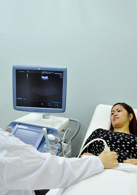
During an ultrasound test, you lie on a table. A special technician or doctor moves a device called a transducer over part of your body. The transducer sends out sound waves, which bounce off the tissues inside your body. The transducer also captures the waves that bounce back. The ultrasound machine creates images from the sound waves.
The images produced by an ultrasound machine can provide valuable information for diagnosing and treating a variety of diseases and conditions.
Most ultrasound examinations are done using a sonar device outside your body, though some ultrasound examinations involve placing a device inside your body.
Ultrasound is used for a variety of reasons, including to:
- View the uterus and ovaries of a pregnant woman and assess her fetus
- Diagnose gallbladder disease
- Evaluate flow in blood vessels
- Guide a needle for biopsy or tumor treatment
- Evaluate a breast lump
- Check a thyroid gland
- Diagnose some cancers
- Reveal genital and prostate abnormalities
Diagnostic ultrasound is a safe procedure that uses low-power sound waves. There are no known risks.
Although ultrasound is a valuable tool, it has limitations. Sound doesn’t travel well through air or bone, so ultrasound isn’t effective at imaging parts of your body that have gas in them or are hidden by bone. To view these areas, your doctor may order other imaging tests, such as CT or MRI scans or X-rays.
Most ultrasound exams require no preparation, with a few exceptions:
- For some ultrasound exams, such as of the gallbladder, your doctor may ask that you not eat or drink for up to 6 hours before the exam.
- Other ultrasound exams, such as of the pelvis, may require a full bladder, so your doctor might ask you to drink up to six glasses of water two hours before the exam and not urinate until the exam is completed.
When scheduling your ultrasound, ask your doctor for specific instructions for your exam.
During an ultrasound exam, you may need to remove jewelry and some or all of your clothing, change into a gown, and lie on an examination table. Gel is applied to your skin to keep air pockets that can block the sound waves from forming.
A trained physician presses a small, hand-held device (transducer), about the size of a bar of soap, against your skin over the area being examined, moving it as necessary to capture the image. The transducer sends sound waves into your body, collects sound waves that bounce back and sends them to a computer, which creates the images.
Some ultrasounds are done inside your body. A transducer is attached to a probe that’s inserted into a natural opening in your body. Examples of these exams include:
- Transesophageal echocardiogram. A transducer is inserted into your esophagus, usually with sedation, to obtain heart images.
- Transrectal ultrasound. A transducer is inserted into a man’s rectum to view his prostate.
- Transvaginal ultrasound. A transducer is inserted into a woman’s vagina to view her uterus and ovaries.
Ultrasound is usually painless. However, you may experience mild discomfort as the physician guides the transducer over your body, especially if you’re required to have a full bladder.
A typical ultrasound exam takes from 30 minutes to an hour.
When your exam is complete, the physician analyzes the images and sends a report to your doctor. Your doctor will share the results with you.
You should be able to return to normal activities immediately after an ultrasound.
What is Ultrasound Imaging of the Abdomen?
An abdominal ultrasound produces a picture of the organs and other structures in the upper abdomen.
A Doppler ultrasound study may be part of an abdominal ultrasound examination.
Doppler ultrasound is a special ultrasound technique that allows the physician to see and evaluate blood flow through arteries and veins in the abdomen, arms, legs, neck and/or brain (in infants and children) or within various body organs such as the liver or kidneys.
What are some common uses of the procedure?
Abdominal ultrasound imaging is performed to evaluate the:
- Kidneys
- Liver
- Gallbladder
- Pancreas
- Spleen
- Abdominal aorta and other blood vessels of the abdomen
Ultrasound is used to help diagnose a variety of conditions, such as:
- Abdominal pain or distention.
- Abnormal liver function.
- Enlarged abdominal organ.
- Stones in the gallbladder or kidney.
- An aneurysm in the aorta.
Additionally, ultrasound may be used to provide guidance for biopsies.
Doppler ultrasound images can help the physician to see and evaluate:
- Blockages to blood flow (such as clots)
- Narrowing of vessels
- Tumors and congenital vascular malformations
- Less than normal or absent blood flow to various organs
- Greater than normal blood flow to different areas which is sometimes seen in infections
What Is Carotid Ultrasound?
Carotid (ka-ROT-id) ultrasound is a painless and harmless test that uses high-frequency sound waves to create pictures of the insides of your carotid arteries.
You have two common carotid arteries, one on each side of your neck. They each divide into internal and external carotid arteries.
The internal carotid arteries supply oxygen-rich blood to your brain. The external carotid arteries supply oxygen-rich blood to your face, scalp, and neck.
Overview
Carotid ultrasound shows whether a waxy substance called plaque (plak) has built up in your carotid arteries. The buildup of plaque in the carotid arteries is called carotid artery disease.
Carotid Arteries
Figure A shows where the right carotid artery is located. Figure B shows a normal carotid artery that has normal blood flow. Figure C shows a carotid artery that has plaque buildup and reduced blood flow.
Over time, plaque can harden or rupture (break open). Hardened plaque narrows the carotid arteries and reduces the flow of oxygen-rich blood to the brain.
If the plaque ruptures, a blood clot can form on its surface. A clot can mostly or completely block blood flow through a carotid artery, which can cause a stroke.
A piece of plaque or a blood clot also can break away from the wall of the carotid artery. The plaque or clot can travel through the bloodstream and get stuck in one of the brain’s smaller arteries. This can block blood flow in the artery and cause a stroke.
A standard carotid ultrasound shows the structure of your carotid arteries. Your carotid ultrasound test might include a Doppler ultrasound. Doppler ultrasound is a special test that shows the movement of blood through your blood vessels.
Your doctor might need results from both types of ultrasound to fully assess whether you have a blood flow problem in your carotid arteries.
What is cranial ultrasound?
Head and transcranial Doppler are two types of cranial ultrasound exams used to evaluate brain tissue and the flow of blood to the brain, respectively.
Head Ultrasound
A head ultrasound examination produces images of the brain and the cerebrospinal fluid that flows and is contained within its ventricles, the fluid filled cavities located in the deep portion of the brain. Since ultrasound waves do not pass through bone easily, this exam is most commonly performed on infants, whose skulls have not completely formed. The gaps between those skull bones provide a “window,” allowing the ultrasound beam to freely pass into and back from the brain. The ultrasound probe and some gel are placed on the outside of the head in one of those regions without bone.
Transcranial Doppler
A transcranial Doppler (TCD) ultrasound evaluates both the direction and velocity of the blood flow in the major cerebral arteries of the brain. This type of ultrasound exam is also used during surgical procedures to monitor blood flow in the brain. TCD may be used alone or with other diagnostic exams such as magnetic resonance imaging (MRI), magnetic resonance angiography (MRA) and computed tomography (CT) scans.
What are some common uses of the procedure?
Head ultrasound is a routine exam for infants who were born prematurely. The procedure is used to screen for brain conditions associated with prematurity, such as bleeding or brain tissue damage as described below. If detected, follow-up ultrasound exams will be performed.
In infants, head ultrasound is used to:
- Evaluate for hydrocephalus, or an enlargement of the ventricles, a condition that can have a number of causes.
- Detect bleeding within the brain tissue or the ventricles. The latter condition is called intraventricular hemorrhage (IVH).
- Assess whether there is damage to the white matter brain tissue surrounding the edges of the ventricles, a condition known as periventricular leukomalacia (PVL).
- Evaluate for congenital abnormalities.
- Locate the site of an infection or tumor.
In adults, head ultrasound is used to locate and evaluate tumor masses during brain surgery, facilitating their safe removal.
Transcranial Doppler ultrasound is used to assess the risk of stroke in adults and children with sickle cell disease. It is also used to measure conditions affecting blood flow to and within the brain, such as:
- Stenosis: a narrowing of a segment of a vessel, most commonly due to atherosclerosis (hardening of the arteries).
- Vasospasm: a temporary narrowing of a vessel, usually a reaction to blood being present in the spinal fluid spaces surrounding the brain. This condition is known as subarachnoid hemorrhage (SAH).
What is a Doppler ultrasound?
A Doppler ultrasound is a noninvasive test that can be used to estimate your blood flow through blood vessels by bouncing high-frequency sound waves (ultrasound) off circulating red blood cells. A regular ultrasound uses sound waves to produce images, but can’t show blood flow.
A Doppler ultrasound may help diagnose many conditions, including:
- Blood clots
- Poorly functioning valves in your leg veins, which can cause blood or other fluids to pool in your legs (venous insufficiency)
- Heart valve defects and congenital heart disease
- A blocked artery (arterial occlusion)
- Decreased blood circulation into your legs (peripheral artery disease)
- Bulging arteries (aneurysms)
- Narrowing of an artery, such as in your neck (carotid artery stenosis)
A Doppler ultrasound can estimate how fast blood flows by measuring the rate of change in its pitch (frequency).
This test may be done as an alternative to more invasive procedures, such as arteriography and venography, which involve injecting dye into the blood vessels so that they show up clearly on X-ray images.
A Doppler ultrasound test may also help your doctor check for injuries to your arteries or to monitor certain treatments to your veins and arteries.
- Echocardiography (EK-o-kar-de-OG-rah-fee), or echo, is a painless test that uses sound waves to create moving pictures of your heart. The pictures show the size and shape of your heart. They also show how well your heart’s chambers and valves are working.
- Echo also can pinpoint areas of heart muscle that aren’t contracting well because of poor blood flow or injury from a previous heart attack. A type of echo called Doppler ultrasound shows how well blood flows through your heart’s chambers and valves.
- Echo can detect possible blood clots inside the heart, fluid buildup in the pericardium (the sac around the heart), and problems with the aorta. The aorta is the main artery that carries oxygen-rich blood from your heart to your body.
- Doctors also use echo to detect heart problems in infants and children.
A fetal ultrasound, or sonogram, is an imaging technique that uses high-frequency sound waves to produce images of a baby in the uterus.
Fetal ultrasound images can help your health care provider evaluate your baby’s growth and development and determine how your pregnancy is progressing. In some cases, fetal ultrasound is used to evaluate possible problems or help confirm a diagnosis.
The first fetal ultrasound is usually done during the first trimester to confirm and date the pregnancy. If your pregnancy remains uncomplicated, the next ultrasound is typically offered during the second trimester, when anatomic details are visible. If your baby’s health needs to be monitored more closely, additional ultrasounds might be recommended.
First trimester ultrasound examination is done to evaluate the presence, size, number and location of the pregnancy. Ultrasound can also be used for first-trimester sonographic genetic screening, as well as screening for any abnormalities of your uterus or cervix.
In the second or third trimester a standard ultrasound is done to evaluate several features of the pregnancy, including fetal anatomy. This exam is typically done between weeks 18 and 20 of pregnancy. However, the timing of this ultrasound might be altered for reasons such as obesity or prior surgical incision at the scanning site, which could limit visualization of the fetus.
During the second and third trimesters, limited ultrasound evaluation might be needed when a specific question requires investigation. Examples include the evaluation of fetal growth and the estimation of amniotic fluid volume. A specialized or detailed exam is done when an anomaly is suspected based on your history or other prenatal exam results.
Here are some of the reasons your health care provider might use fetal ultrasound:
- Confirm the pregnancy and its location. Some fetuses develop outside of the uterus, in the fallopian tube. A fetal ultrasound can help your health care provider detect a pregnancy outside of the uterus (ectopic pregnancy).
- Determine your baby’s gestational age. Knowing the baby’s age can help your health care provider determine your due date and track various milestones throughout your pregnancy.
- Confirm the number of babies. If your health care provider suspects a multiple pregnancy, an ultrasound might be done to confirm the number of babies.
- Evaluate your baby’s growth. Your health care provider can use ultrasound to determine whether your baby is growing at a normal rate. Ultrasound can be used to monitor your baby’s movement, breathing and heart rate.
- Study the placenta and amniotic fluid levels. The placenta provides your baby with vital nutrients and oxygen-rich blood. Too much or too little amniotic fluid — the fluid that surrounds the baby in the uterus during pregnancy — or complications with the placenta need special attention. An ultrasound can help evaluate the placenta and amniotic fluid around the baby.
- Identify birth defects. An ultrasound can help your health care provider screen for some birth defects.
- Investigate complications. If you’re bleeding or having other complications, an ultrasound might help your health care provider determine the cause.
- Perform other prenatal tests. Your health care provider might use ultrasound to guide needle placement during certain prenatal tests, such as amniocentesis or chorionic villus sampling.
- Determine fetal position before delivery. Most babies are positioned headfirst by the end of the third trimester. That doesn’t always happen, though. Ultrasound imaging can confirm the baby’s presentation so that your health care provider can discuss options for delivery.
Fetal ultrasound should be done only for valid medical reasons. Fetal ultrasound isn’t recommended simply to determine a baby’s sex. Similarly, fetal ultrasound isn’t recommended solely for the purpose of producing keepsake videos or pictures.
If your health care provider doesn’t suggest a fetal ultrasound but you’d like the reassurance an ultrasound can provide, share your wishes with your provider so that you can work together to determine what’s best for you and your baby.
Diagnostic ultrasound has been used during pregnancy for many years and is generally considered safe when used appropriately. The lowest amount of ultrasound energy that provides an accurate assessment should be used. Obstetric providers and ultrasonographers are trained in appropriate use of ultrasound energy.
Ultrasound should be requested only by a doctor or licensed health care provider for medical reasons. The use of fetal ultrasound solely to create keepsake photos or videos isn’t recommended.
Fetal ultrasound also has limitations. Fetal ultrasound might not detect all birth defects — or might incorrectly suggest a birth defect is present when it’s not.
You might be asked to drink a certain amount of fluid or avoid urinating before a fetal ultrasound, depending on the type of ultrasound. When scheduling your ultrasound, ask your health care provider for specific instructions.
Also be aware that fetal ultrasound can be done through the vagina (transvaginal) or over the abdomen (transabdominal), depending on why it’s being done or the stage of your pregnancy. If you’re having a transabdominal ultrasound, consider wearing loose fitting clothing so that you can easily expose your abdomen.
There are two main types of fetal ultrasound exams:
- Transvaginal ultrasound. With this type of fetal ultrasound, a wandlike transducer is placed in your vagina to send out sound waves and gather the reflections. Transvaginal ultrasounds are used most often during early pregnancy, when the uterus and fallopian tubes are closer to the vagina than to the abdominal surface.
- Transabdominal ultrasound. A transabdominal fetal ultrasound is done by moving a transducer — a small plastic device that sends and receives sound waves — over your abdomen. This type of fetal ultrasound is usually used during the later part of your pregnancy.
Various other types of transabdominal ultrasounds are available, including:
- Specialized sonographic evaluation. Specialized examination might be needed in specific circumstances, such as when a fetal abnormality is known or suspected. In this situation, a more detailed evaluation can provide additional information about the abnormality.
- 4-D ultrasound. A 4-D fetal ultrasound can provide images of a baby with photo-quality details. This type of ultrasound is sometimes used to help health care providers detect facial abnormalities or neural tube defects.
- Doppler ultrasound. A Doppler ultrasound measures slight changes in the ultrasound waves as they bounce off moving objects, such as blood cells. A Doppler ultrasound can provide details about a baby’s blood flow.
- Fetal echocardiography. This type of fetal ultrasound provides a detailed picture of a baby’s heart. It might be used to confirm or rule out a congenital heart defect.
During the exam
During a transabdominal fetal ultrasound, you’ll recline on an exam table and expose your abdomen. Your health care provider will apply a special gel to your abdomen. This will improve the conduction of sound waves and eliminate air between your skin and the transducer — the small plastic device that sends out sound waves and receives those that bounce back.
Your health care provider will move the transducer back and forth over your abdomen. The sound waves reflected off your bones and other tissues will be converted into images on a monitor.
Your health care provider will measure your baby’s anatomy. He or she might print or store certain images to document important structures. You’ll likely be given copies of some of the images.
Depending on your baby’s position and stage of development, you might be able to make out a face, hands and fingers, or arms and legs. Don’t worry if you can’t “see” your baby. Ultrasound images can be hard for an untrained observer to decipher.
What is Ultrasound Imaging of the Hip?
Ultrasound images of the hip provide pictures of muscles, tendons, ligaments, joints, bone and soft tissues of the hip. In infants, the hip (which has a ball and cup configuration) is composed mainly of cartilage and is easily recognized on ultrasound.
What are some common uses of the procedure?
Hip ultrasound images are typically used to help evaluate:
- Abnormalities of the muscles, such as tears and soft-tissue masses.
- Foreign bodies, bleeding, infections or other types of fluid collections.
- Benign and malignant soft tissue tumors.
- Early changes of arthritis.
- Infant ultrasound can be used to check the hips for developmental dysplasia of the hip (DDH), which in infants can range from a shallow cup (bony acetabular dysplasia), to complete dislocation with the ball of the femoral head completely outside the socket.
What is Ultrasound Imaging of the Musculoskeletal System?
Ultrasound images of the musculoskeletal system provide pictures of muscles, tendons, ligaments, joints and soft tissue throughout the body.
What are some common uses of the procedure?
Ultrasound images are typically used to help diagnose:
- Tendon tears, or tendinitis of the rotator cuff in the shoulder, Achilles tendon in the ankle and other tendons throughout the body.
- Muscle tears, masses or fluid collections.
- Ligament sprains or tears.
- Inflammation or fluid (effusions) within the bursae and joints.
- Early changes of rheumatoid arthritis.
- Nerve entrapments such as carpal tunnel syndrome.
- Benign and malignant soft tissue tumors.
- Ganglion cysts.
- Hernias.
- Foreign bodies in the soft tissues (such as splinters or glass).
- Dislocations of the hip in infants.
- Fluid in a painful hip joint in children.
- Neck muscle abnormalities in infants with torticollis (neck twisting).
- Soft tissue masses (lumps/bumps) in children.
What is Pelvic Ultrasound Imaging?
There are three types of pelvic ultrasound:
- Abdominal (transabdominal)
- Vaginal (transvaginal, endovaginal) for women
- Rectal (transrectal) for men
A Doppler ultrasound exam may be part of a pelvic ultrasound examination.
Doppler ultrasound is a special ultrasound technique that allows the physician to see and evaluate blood flow through arteries and veins in the abdomen, arms, legs, neck and/or brain (in infants and children) or within various body organs such as the liver or kidneys.
What are some common uses of the procedure?
In women, a pelvic ultrasound is most often performed to evaluate the:
- Uterus
- Cervix
- Ovaries
- Fallopian tubes
- Bladder
Pelvic ultrasound exams are also used to monitor the health and development of an embryo or fetus during pregnancy. See theObstetrical Ultrasound page for more information.
Ultrasound examinations can help diagnose symptoms experienced by women such as:
- Pelvic pain
- Abnormal bleeding
- Other menstrual problems and help identify:
- Palpable masses such as ovarian cysts and uterine fibroids
- Ovarian or uterine cancers
A transvaginal ultrasound is usually performed to view the endometrium, the lining of the uterus, and the ovaries. Transvaginal ultrasound also provides a good way to evaluate the muscular walls of the uterus, called the myometrium.
Sonohysterography allows for a more in-depth investigation of the uterine cavity. Three-dimensional (3-D) ultrasound permits evaluation of the uterus and ovaries in planes that cannot be imaged directly. These exams are typically performed to detect:
- Uterine anomalies
- Uterine scars
- Endometrial polyps
- Fibroids
- Cancer, especially in patients with abnormal uterine bleeding
Some physicians also use 3-D ultrasound or sonohysterography for patients with infertility. Three-dimensional ultrasound provides information about the outer contour of the uterus and about uterine irregularities. See the Sonohysterography page for more information.
In men, a pelvic ultrasound is used to evaluate the:
- Bladder
- Seminal vesicles
- Prostate
Transrectal ultrasound, a special study usually done to view the prostate gland, involves inserting a specialized ultrasound transducer into a man’s rectum. See the Prostate Ultrasound page for more information.
In men and women, a pelvic ultrasound exam can help identify:
- Kidney stones
- Bladder tumors
- Other disorders of the urinary bladder
In children, pelvic ultrasound can help evaluate:
- Pelvic masses
- Pelvic pain
- Ambiguous genitalia and anomalies of pelvic organs
- Early or delayed puberty in girls
Pelvic ultrasound is also used to guide procedures such as needle biopsies, in which needles are used to extract a sample of cells from organs for laboratory testing.
Doppler ultrasound images can help the physician to see and evaluate:
- Blockages to blood flow (such as clots)
- Narrowing of vessels
- Tumors and congenital vascular malformations
- Less than normal or absent blood flow to various organs
- Greater than normal blood flow to different areas which is sometimes seen in infections
What is Vascular Ultrasound?
Vascular ultrasound provides pictures of the body’s veins and arteries.
A Doppler ultrasound study is usually part of a vascular ultrasound examination.
Doppler ultrasound is a special ultrasound technique that allows the physician to see and evaluate blood flow through arteries and veins in the abdomen, arms, legs, neck and/or brain (in infants and children) or within various body organs such as the liver or kidneys.
What are some common uses of the procedure?
Sonography is a useful way of evaluating the body’s circulatory system. Vascular ultrasound is performed to:
- Help monitor the blood flow to organs and tissues throughout the body.
- Locate and identify blockages (stenosis) and abnormalities like plaque or emboli and help plan for their effective treatment.
- Detect blood clots (deep venous thrombosis (DVT) in the major veins of the legs or arms.
- Determine whether a patient is a good candidate for a procedure such as angioplasty.
- Evaluate the success of procedures that graft or bypass blood vessels.
- Determine if there is an enlarged artery (aneurysm).
- Determine the source and severity of varicose veins.
In children, ultrasound is used to: - Aid in the placement of a needle or catheter into a vein or artery to help avoid complications such as bleeding, nerve injury or pseudo-aneurysm (abnormal outpouching of an artery with the risk of rupture).
- Evaluate a connection between an artery and a vein which can be seen in congenital vascular malformations (arteriovenous malformations or fistula) and in dialysis fistula.
If a line is placed in an artery or vein of the legs or arms, there is a much higher chance of developing a clot around it due to the smaller vessel size (especially in infants and young children). In some instances, a clot can form in the arm or in the left leg with the latter extending into the major vein of the abdomen. Plaque formation is not frequently seen in children but there can be compression at the inlet of the chest.
Doppler ultrasound images can help the physician to see and evaluate:
- Blockages to blood flow (such as clots)
- Narrowing of vessels
- Tumors and congenital vascular malformations
- Less than normal or absent blood flow to various organs
- Greater than normal blood flow to different areas which is sometimes seen in infections