Diagnostic ultrasound, also called sonography, is a type of imaging. It uses high-frequency sound waves to look at organs and structures inside the body. Health care professionals use it to view the heart, blood vessels, kidneys, liver, and other organs. During pregnancy, doctors use ultrasound to view the fetus. Unlike x-rays, ultrasound does not expose you to radiation.
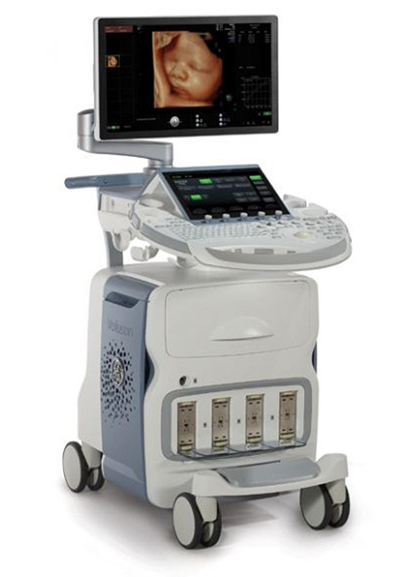
During an ultrasound test, you lie on a table. A special technician or doctor moves a device called a transducer over part of your body. The transducer sends out sound waves, which bounce off the tissues inside your body. The transducer also captures the waves that bounce back. The ultrasound machine creates images from the sound waves.
The images produced by an ultrasound machine can provide valuable information for diagnosing and treating a variety of diseases and conditions.
Most ultrasound examinations are done using a sonar device outside your body, though some ultrasound examinations involve placing a device inside your body.
Ultrasound is used for a variety of reasons, including to:
- View the uterus and ovaries of a pregnant woman and assess her fetus
- Diagnose gallbladder disease
- Evaluate flow in blood vessels
- Guide a needle for biopsy or tumor treatment
- Evaluate a breast lump
- Check a thyroid gland
- Diagnose some cancers
- Reveal genital and prostate abnormalities
Diagnostic ultrasound is a safe procedure that uses low-power sound waves. There are no known risks.
Although ultrasound is a valuable tool, it has limitations. Sound doesn’t travel well through air or bone, so ultrasound isn’t effective at imaging parts of your body that have gas in them or are hidden by bone. To view these areas, your doctor may order other imaging tests, such as CT or MRI scans or X-rays.
Most ultrasound exams require no preparation, with a few exceptions:
- For some ultrasound exams, such as of the gallbladder, your doctor may ask that you not eat or drink for up to 6 hours before the exam.
- Other ultrasound exams, such as of the pelvis, may require a full bladder, so your doctor might ask you to drink up to six glasses of water two hours before the exam and not urinate until the exam is completed.
When scheduling your ultrasound, ask your doctor for specific instructions for your exam.
During an ultrasound exam, you may need to remove jewelry and some or all of your clothing, change into a gown, and lie on an examination table. Gel is applied to your skin to keep air pockets that can block the sound waves from forming.
A trained physician presses a small, hand-held device (transducer), about the size of a bar of soap, against your skin over the area being examined, moving it as necessary to capture the image. The transducer sends sound waves into your body, collects sound waves that bounce back and sends them to a computer, which creates the images.
A fetal ultrasound, or sonogram, is an imaging technique that uses high-frequency sound waves to produce images of a baby in the uterus.
Fetal ultrasound images can help your health care provider evaluate your baby’s growth and development and determine how your pregnancy is progressing. In some cases, fetal ultrasound is used to evaluate possible problems or help confirm a diagnosis.
The first fetal ultrasound is usually done during the first trimester to confirm and date the pregnancy. If your pregnancy remains uncomplicated, the next ultrasound is typically offered during the second trimester, when anatomic details are visible. If your baby’s health needs to be monitored more closely, additional ultrasounds might be recommended.
First trimester ultrasound examination is done to evaluate the presence, size, number and location of the pregnancy. Ultrasound can also be used for first-trimester sonographic genetic screening, as well as screening for any abnormalities of your uterus or cervix.
In the second or third trimester a standard ultrasound is done to evaluate several features of the pregnancy, including fetal anatomy. This exam is typically done between weeks 18 and 20 of pregnancy. However, the timing of this ultrasound might be altered for reasons such as obesity or prior surgical incision at the scanning site, which could limit visualization of the fetus.
During the second and third trimesters, limited ultrasound evaluation might be needed when a specific question requires investigation. Examples include the evaluation of fetal growth and the estimation of amniotic fluid volume. A specialized or detailed exam is done when an anomaly is suspected based on your history or other prenatal exam results.
Here are some of the reasons your health care provider might use fetal ultrasound:
- Confirm the pregnancy and its location. Some fetuses develop outside of the uterus, in the fallopian tube. A fetal ultrasound can help your health care provider detect a pregnancy outside of the uterus (ectopic pregnancy).
- Determine your baby’s gestational age. Knowing the baby’s age can help your health care provider determine your due date and track various milestones throughout your pregnancy.
- Confirm the number of babies. If your health care provider suspects a multiple pregnancy, an ultrasound might be done to confirm the number of babies.
- Evaluate your baby’s growth. Your health care provider can use ultrasound to determine whether your baby is growing at a normal rate. Ultrasound can be used to monitor your baby’s movement, breathing and heart rate.
- Study the placenta and amniotic fluid levels. The placenta provides your baby with vital nutrients and oxygen-rich blood. Too much or too little amniotic fluid — the fluid that surrounds the baby in the uterus during pregnancy — or complications with the placenta need special attention. An ultrasound can help evaluate the placenta and amniotic fluid around the baby.
- Identify birth defects. An ultrasound can help your health care provider screen for some birth defects.
- Investigate complications. If you’re bleeding or having other complications, an ultrasound might help your health care provider determine the cause.
- Perform other prenatal tests. Your health care provider might use ultrasound to guide needle placement during certain prenatal tests, such as amniocentesis or chorionic villus sampling.
- Determine fetal position before delivery. Most babies are positioned headfirst by the end of the third trimester. That doesn’t always happen, though. Ultrasound imaging can confirm the baby’s presentation so that your health care provider can discuss options for delivery.
Fetal ultrasound should be done only for valid medical reasons. Fetal ultrasound isn’t recommended simply to determine a baby’s sex. Similarly, fetal ultrasound isn’t recommended solely for the purpose of producing keepsake videos or pictures.
If your health care provider doesn’t suggest a fetal ultrasound but you’d like the reassurance an ultrasound can provide, share your wishes with your provider so that you can work together to determine what’s best for you and your baby.
Diagnostic ultrasound has been used during pregnancy for many years and is generally considered safe when used appropriately. The lowest amount of ultrasound energy that provides an accurate assessment should be used. Obstetric providers and ultrasonographers are trained in appropriate use of ultrasound energy.
Ultrasound should be requested only by a doctor or licensed health care provider for medical reasons. The use of fetal ultrasound solely to create keepsake photos or videos isn’t recommended.
Fetal ultrasound also has limitations. Fetal ultrasound might not detect all birth defects — or might incorrectly suggest a birth defect is present when it’s not.
You might be asked to drink a certain amount of fluid or avoid urinating before a fetal ultrasound, depending on the type of ultrasound. When scheduling your ultrasound, ask your health care provider for specific instructions.
Also be aware that fetal ultrasound can be done through the vagina (transvaginal) or over the abdomen (transabdominal), depending on why it’s being done or the stage of your pregnancy. If you’re having a transabdominal ultrasound, consider wearing loose fitting clothing so that you can easily expose your abdomen.
There are two main types of fetal ultrasound exams:
- Transvaginal ultrasound. With this type of fetal ultrasound, a wandlike transducer is placed in your vagina to send out sound waves and gather the reflections. Transvaginal ultrasounds are used most often during early pregnancy, when the uterus and fallopian tubes are closer to the vagina than to the abdominal surface.
- Transabdominal ultrasound. A transabdominal fetal ultrasound is done by moving a transducer — a small plastic device that sends and receives sound waves — over your abdomen. This type of fetal ultrasound is usually used during the later part of your pregnancy.
Various other types of transabdominal ultrasounds are available, including:
- Specialized sonographic evaluation. Specialized examination might be needed in specific circumstances, such as when a fetal abnormality is known or suspected. In this situation, a more detailed evaluation can provide additional information about the abnormality.
- 4-D ultrasound. A 4-D fetal ultrasound can provide images of a baby with photo-quality details. This type of ultrasound is sometimes used to help health care providers detect facial abnormalities or neural tube defects.
- Doppler ultrasound. A Doppler ultrasound measures slight changes in the ultrasound waves as they bounce off moving objects, such as blood cells. A Doppler ultrasound can provide details about a baby’s blood flow.
- Fetal echocardiography. This type of fetal ultrasound provides a detailed picture of a baby’s heart. It might be used to confirm or rule out a congenital heart defect.
During the exam
During a transabdominal fetal ultrasound, you’ll recline on an exam table and expose your abdomen. Your health care provider will apply a special gel to your abdomen. This will improve the conduction of sound waves and eliminate air between your skin and the transducer — the small plastic device that sends out sound waves and receives those that bounce back.
Your health care provider will move the transducer back and forth over your abdomen. The sound waves reflected off your bones and other tissues will be converted into images on a monitor.
Your health care provider will measure your baby’s anatomy. He or she might print or store certain images to document important structures. You’ll likely be given copies of some of the images.
Depending on your baby’s position and stage of development, you might be able to make out a face, hands and fingers, or arms and legs. Don’t worry if you can’t “see” your baby. Ultrasound images can be hard for an untrained observer to decipher.